骨科X线阅片的4个步骤:ABCS,一步一步来看
来源:内固定学习平台
X线片子在骨科*常见,*基础。
拿到一张片子,怎么阅片?是有章法的!今天我们来学习~
X-ray interpretation (ABCS approach)
X 射线解读(ABCS 方法)
TheABCS approach of X-ray interpretation involves assessing the following:
X 线解读的 ABCS 方法主要评估以下内容:
Alignment and joint space
对位对线和关节间隙 Bone texture
骨小梁纹理 Cortices
皮质 Soft tissues
软组织
General points一般要点
Don’t forget to reviewall views, compare both sides and re‐examine any previous imaging.
不要忘记审查所有视图,比较两侧并重新检查任何以前的影像。
If you spot one abnormality,do not lose focus until you have reviewed all areas of the image, otherwise, you might miss important pathology.
如果您发现一个异常,不要失去方寸,直到您已审查完图像的所有区域,否则您可能会错过重要的病理学发现。
Alignment and joint space
对位关系和关节间隙
Changes in alignment suggest afracture, subluxation (partial dislocation) or dislocation.
对位改变提示骨折、半脱位(部分脱位)或脱位。
When describing the displacement, the position of thefragment distal to the fracture site is always described.
描述移位时,总是描述骨折部位远端骨折片的位置。
The radiograph below demonstrates why it is important to havemore than one view.
以下 X 光片展示了为什么需要不止一个视角的重要性。
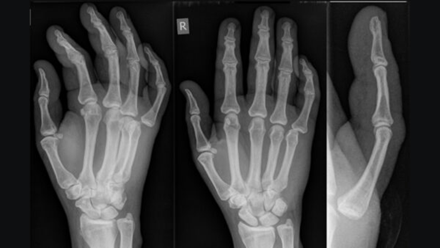
Joint dislocation of the metacarpophalangeal joint of the 5th digit.1
掌指关节第五指关节脱位。
Carefully look at thejoint space to identify changes such as joint space narrowing due to cartilage loss, cartilage calcification (as in chondrocalcinosis) or new bone formation (e.g. osteophytes). Subchondral sclerosis (increased bone density) is often present along the joint lines in patients with osteoarthritis.
仔细观察关节间隙,以识别如因软骨损失导致的关节间隙变窄、软骨钙化(如软骨钙化症)或新骨形成(例如骨刺)等变化。骨关节炎患者关节线附近常伴有软骨下硬化(骨密度增加)。
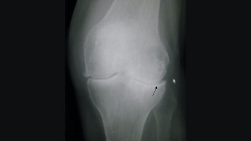
Osteoarthritis of the left knee. Note the osteophytes (right side of joint), narrowing of the joint space, and increased subchondral bone density (black arrow).2
左膝骨关节炎。注意骨刺(关节右侧)、关节间隙变窄和软骨下骨密度增加(黑箭头)。
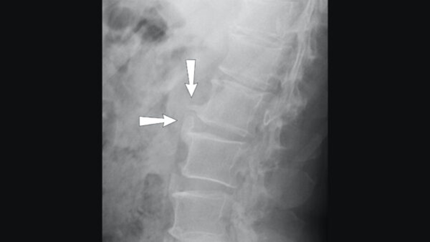
Lateral X-ray of spondylosis of the lumbar spine, with osteophytes marked by arrows3
侧位腰椎椎间盘突出 X 光片,箭头标记骨刺 3
Bone texture骨小梁纹理
Altered density or disruption in the usual internal matrix of fine white lines (trabeculae) within the substance of the bone and the thick external covering (cortex) may indicate pathology.
骨实质内细白线(骨小梁)的密度改变或正常内部结构的破坏,以及厚厚的外部覆盖层(皮质)的破坏,可能表明存在病理情况。
 Healthy cortex and trabeculae vs osteomyelitis.4,5
健康皮质和小梁与骨髓炎。
Cortices皮质
Trace around the outline of each bone as anystep in the cortex may indicate a fracture or other pathology.
围绕每块骨骼的轮廓进行追踪,因为皮质中的任何一步都可能表明骨折或其他病理情况。
Infection and tumours (primary and secondary) are the commonest causes of bony destruction.
感染和肿瘤(原发性和继发性)是骨破坏*常见的原因。
A periosteal reaction is the formation of new bone in response to injury or other stimuli of the periosteum surrounding the bone. It may be the only sign visible to denote a problem with the bone (stress or healing fracture, mild osteomyelitis or tumour).
骨膜反应是指骨膜周围受到损伤或其他刺激后新骨的形成。它可能是**可见的迹象,表明存在骨骼问题(应力性骨折或愈合骨折、轻度骨髓炎或肿瘤)。
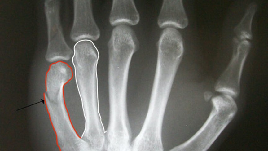
Trace around the bone cortex carefully to identify fractures (arrow)6
仔细沿骨皮质周围追踪以识别骨折(箭头) 6
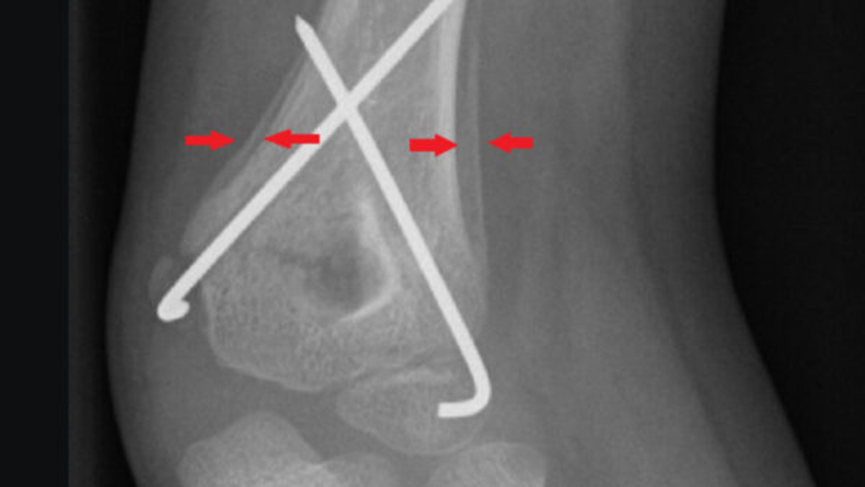
Periosteal reaction by a supracondylar fracture7
骨膜反应,肱骨髁上骨折 7
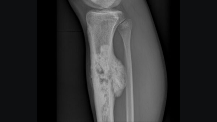
Osteosarcoma affecting the tibia8
骨肉瘤累及胫骨
Soft tissues软组织
Look for anyswelling, foreign bodies or effusions. Sometimes soft tissue injuries are easier to visualise and can prompt a closer inspection for bony pathology.
寻找任何肿胀、异物或渗出物。有时软组织损伤更容易可视化,可以促使更仔细地检查骨骼病理学。
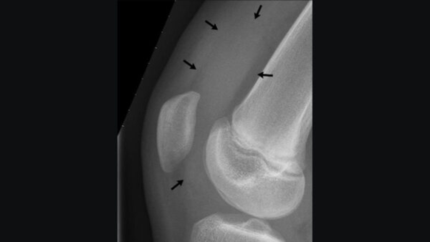
X-ray of the knee of a 12-year-old male, showing knee effusion of medium severity, marked by black arrows. It displaces the patella anteriorly and extends into the suprapatellar bursa.9
膝关节 12 岁男孩 X 光片,显示中等程度膝关节积液,以黑箭头标记。使髌骨向前移位并延伸至髌上囊。 9
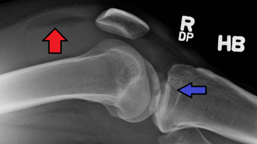
Lipohaemarthrosis (red arrow) due to a tibial plateau fracture (blue arrow)10
脂血性关节积血(红箭头)由胫骨平台骨折(蓝箭头)引起 10